
Melittin is 40 – 50% of dried bee venom composition[1]
[1] Cancer Lett. 2017 doi: 10.1016/j.canlet.2017.05.010 PMCID: PMC5682937 NIHMSID: NIHMS915401
 Polypeptide Melittin – major hemolytic component of Bee Venom.
- Chemical Names: Melitten; Mellitin; Forapine; Melittin (honeybee), etc. Contains 26 amino acids, has cytolytic properties, causes contracture of muscle, releases histamine, and disrupts surface tension, probably due to lysis of cell and mitochondrial membranes.
- Production technology:
- organic – purified from the bee venom
-
synthetic – chemical synthesis
-
Empirical Formula (Hill Notation): C131H229N39O31
-
Molecular Weight and structure:[1]
-
Monomeric form 2,846 Daltons;
-
Tetrameric form 12,500 Daltons
-
-
CAS RN: 20449-79-0 or 37231-28-0; CHEBI: 6736; PubChem CID: 16133648
-
MeSH Descriptor Data: [D12.644.050.550]; FDA UNII: 24VT8NVE75
[1] Terwilliger TC, Eisenberg D. The structure of melittin. I. Structure determination and partial refinement. J. Biol. Chem. (1982) https://www.ncbi.nlm.nih.gov/Structure/pdb/2MLT
Early studies of AMPs relied on purifying peptides from natural sources and assessing the antibacterial activity of these extracted samples. Chemical synthesis of peptides has quickly become the preferred method for obtaining AMPs at high yield and purity. Solid-phase peptide synthesis is widely used to generate large quantities of peptide.[1] In addition to synthetic peptides, many research groups have used molecular cloning technology to recombinantly express and purify AMPs. Another strategy involves genetically engineering eukaryotic yeast cells, such as Pichia pastoris, to produce the AMPs of interest.[2]
Comparative studies of native and synthetic melittins[3].
The hexacosapeptide melittin-I, has been synthesized by Lübke and Schröder. In addition, the following derivatives have been prepared which are probably also present in bee venom: melittin II (which differs by one serine), and N1-formylated melittin-I and II.
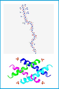
The structure, forms and conformations[1],[2],[3]
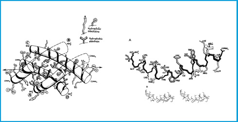
Melittin can adopt a variety of conformations in different media: in water it exists as a random-coil monomer or an A-helical tetramer depending on concentration, pH and ionic strength, and it is a largely helical monomer in organic solvents or in the presence of detergent micelles or vesicles. There is a crystal structure of the tetramer, which is made up of a dimer of dimers. It is likely that on contact with membranes, melittin initially forms an amphipathic helix on the membrane surface. To do this, it has to dissociate from the tetrameric state that it has in solution.
MOLBASE is one of the world’s largest integrated platforms for chemical e-commerce, enabling industry professionals to discover, evaluate, and acquire chemicals in a more transparent, efficient, and convenient fashion.
http://www.molbase.com/en/search.html?search_keyword=melittin
- As part of the innate immune system, AMPs (AntiMicrobial Peptides) are defensive peptides with no antimicrobial resistance, that efficiently penetrate infected cells and tissues beyond many endothelial barriers, most linings and overall specifically target pathogens.
- The therapeutic value of Honey Bee Venom (BV) has been acknowledged for over a hundreds years. Subsequently, the application scope expanded from conventional antinociceptive effect (reduced sensitivity to pain) to degenerative diseases of the nervous system and immunomodulatory effects.
- Are recognized the therapeutic effects of Melittin against inflammatory and parasitic diseases, including skin inflammation, Neuro inflammation, Arthritis, Atherosclerosis and liver inflammation, inhibition of the pathogen biofilm formations like Staphylococcus aureus, Escherichia coli and Pseudomonas aeruginosa.
- Melittin is perspective in destruction the cancer cells.
- Pharmacological synergism of melittin with antibiotics and plant secondary metabolites against multi-drug resistant microbial pathogens.
- Other therapeutic properties of melittin, such as anti-HIV prospects, immunotherapy, the blood anti-coagulation, transport of drugs into the cell, etc. are investigated.
There are thousands of publications (PubMed, Springer, ResearchGate, etc.) and patents[1],[2] devoted to melittin prospect applications and its isolation methods. However, to date none of these have achieved new drug approval [3]. One main drawback to marketing melittin is its high cost.
Some therapeutic applications of bee venom (BV) and its components [4]
|
Disease Type |
Component |
|
Parkinson’s disease |
Bee venom (BV) |
|
Apamin |
|
|
BV |
|
|
Amyotrophic lateral sclerosis (ALS) |
BV |
|
Melittin |
|
|
BV |
|
|
Multiple sclerosis |
BV |
|
Cancer |
BV |
|
Melittin |
|
|
Liver fibrosis |
BV |
|
Apamin |
|
|
Melittin |
|
|
PLA2 |
|
|
Atherosclerosis |
BV |
|
Apamin |
|
|
Melittin |
|
|
Skin disease (acne vulgaris) |
BV |
|
Melittin |
|
|
Skin disease (atopic dermatitis) |
BV |
|
Learning deficit |
Apamin |
|
Pain |
BV |
|
|
BV |
|
Lupus nephritis |
BV |
Write-up of main therapeutic and Biotechnological Applications of Melittin with the broad references.[5]
Melittin for Therapeutic Use.
Antimicrobial peptides (AMPs) have been widely studied as an alternative to conventional antibiotics, especially for the treatment of drug-resistant infections. Hundreds of AMPs have been isolated, and several thousand have been de novo designed and synthesized. Despite displaying extensive sequence heterogeneity, most of these peptides share two functionally important features, namely a net positive charge and the ability to adopt an amphipathic structure. Melittin is considered to show strong antimicrobial properties and it also has hemolytic activity and marked allergenic properties. Early studies using individual peptide analogs of melittin showed that the initial step of the mechanism underlying the hemolytic and antimicrobial activity of this venom peptide involves interactions with the lipid groups of the membrane. The structural requirements for the action of melittin, its orientation, aggregation state, current view of pore formation, and also its various cellular actions are discussed in detail in an excellent review by Dempsey. Bruce Merrifield performed pioneering work on improving the features of antimicrobial peptides, shortening their sequences and increasing their activity. In particular, a hybrid undecapeptide derived from the well-known cecropin A and melittin was found to be sufficient for antifungal and antibacterial activities, while displaying low cytotoxicity. This hybrid version was later improved with retro and retroenantio analogs. Indeed, a patent of several active d-peptides with antibiotic and antimalarial activity was even filed. Despite the therapeutic efficacy of antimicrobial peptides, their use is limited due to poor in vivo bioavailability caused by instability, cytotoxicity, hydrophobicity, in addition, the cost production is an issue. In parallel to antimicrobial peptides for therapeutic use in humans, these peptides can be applied to fight economically important plant pathogens, which are currently one of the major factors limiting crop production worldwide. A library of linear undecapeptides derived from cecropin-melittin hybrids have been tested against phytopathogenic bacteria and patented for future use in phytosanitary compositions. In this regard, a promising peptide called BP76 has been identified for this purpose.
Antimicrobial Properties of Melittin for Biotechnological Use
The idea of using antimicrobial peptides has also been translated to coatings for medical devices. Currently, a number of companies are turning their attention to the use of antimicrobial coatings of cationic peptides, such as melittin, for contact lenses in order to prevent the growth of undesirable microorganisms. Contact lenses made of materials comprising hydrogels and antimicrobial ceramics that contain at least one metal (selected from Ag, Cu and Zn) are available. However, although these polymeric compositions do have antimicrobial properties, they do not have all the properties desired for extended-wear contact lenses. Antimicrobial coatings containing covalently bound antimicrobial peptides exhibit diminished activity when compared that of the unbound corresponding antimicrobial peptides in solution. To overcome these drawbacks, Novartis has patented a method to produce contact lenses with an antimicrobial metal-containing layer-by-layer (LbL). In its LbL design, at least one layer has a negatively charged polyionic material, having -COO-Ag groups or silver nanoparticles.
The antiviral activities described for melittin and its analogs are caused by specific intracellular events, with the selective reduction of the biosynthesis of some viral proteins, as reported for the melittin analog Hecate on herpes virus-1, and for melittin itself on HIV-1-infected lymphoma cells. In the 90s, active melittin was presented to provide an improved composition complementary to azidothymidine (AZT) to inhibit the reverse transcriptase and growth of HIV-infected cells. Recently, a similar idea has been patented, whereby melittin is carried in a nanoparticle construct designed to be used as a topical vaginal virucide.
Related to fields of immunology and vaccinology, the 90s also witnessed great progress in therapeutic approaches based on vaccination against infectious pathogens. Despite these advances in the identification of new antigens and their immunological mechanisms, the immune response in most cases continues to be very weak. Therefore, to improve the response, effective adjuvants to enhance the immunogenicity of target antigens must be used. A few years ago, Rinaldo Zurbriggen presented a novel adjuvant system based on melittin and analogs capable of eliciting strong immune responses against target antigens, thus reducing the risk of toxic side effects associated with the use of adjuvants.
Uncontrolled inflammation can cause extensive tissue damage and is the hallmark of numerous diseases, including rheumatoid arthritis, which results in joint destruction and permanent disability. PLA2 is the enzyme responsible for hydrolyzing arachidonic acid from phospholipids, and arachidonic acid is the precursor of eicosanoids, which are thought to mediate inflammation. Melittin and related peptides have been described as anti-inflammatory drugs as they have the capacity to inhibit PLA2. However, in this field, melittin competes with a wide variety of non-steroidal drugs, methotrexate, and other biological disease-modifying antirheumatic drugs.
Bee venom (BV) has been used for millennia in Chinese traditional medicine to treat rheumatoid arthritis (RA) [1]. On the way of development the treatment of RA examined the anti-arthritis effects of melittin on the complete Freund’s adjuvant-induced (CFA-induced) RA model in rats. The RA animal models were treated with solutions of BV, melittin, and saline by injection into a specific acupoint (Zusanli). The BV and melittin treatments statistically diminished the thickness of the arthroses in the injected side of the paw, compared to the saline treatment. Melittin therapy also significantly reduced arthritis-induced nociceptive behaviors, as assessed by the thermal hyperalgesia test. In addition, CFA-induced Fos expression in the superficial layer of the lumbar spinal cord was significantly suppressed by the BV and melittin treatments, compared to the saline treatment. These results indicate that melittin is an effective anti-arthritis component of whole bee venom, making it a promising candidate as an anti-arthritis drug.
[1] Jinghua Li, Tao Ke, Chao He, Wei Cao, Mengqi Wei, Lei Zhang, Jin-Xia Zhang, Wen Wang, Jing Ma, Zong-Ren Wang, and Zhong-Jun Shao. The Anti-Arthritic Effects of Synthetic Melittin on the Complete Freund’s Adjuvant-Induced Rheumatoid Arthritis Model in Rats. The American Journal of Chinese Medicine (2010) DOI: 10.1142/S0192415X10008457
Atherosclerosis
Atherosclerosis is the major cause of morbidity and mortality worldwide. This specific form of arteriosclerosis is a chronic inflammatory disease of the arteries caused by the accumulation and interaction of white blood cells, remnants of dead cells, cholesterol, and triglycerides on the artery wall. This complex inflammatory process is characterized by the presence of monocytes/macrophages and T lymphocytes in the atheroma, where macrophages secrete pro-inflammatory cytokines, a main cellular component in the development of atherosclerotic plaques. Several in vitro studies have shown positive effects of melittin for the treatment of atherosclerosis. In addition, in vivo experiments have demonstrated the molecular mechanism of the anti-atherosclerotic effects of melittin in mouse models of this disease. This has been the major finding regarding the capacity of melittin to prevent lipopolysaccharide (LPS)/high-fat-induced expression of inflammatory cytokines, proatherogenic proteins, and adhesion molecules.
Anti-Cancer
Many studies report that melittin inhibits tumor cell growth and induces apoptosis, thereby indicating a potential application of this venom peptide as an alternative or complementary medicine for the treatment of human cancers. A valuable review describing the mechanisms underlying the anticancer effects of melittin has been published.
Mechanisms of action of cationic antitumor peptides[1]
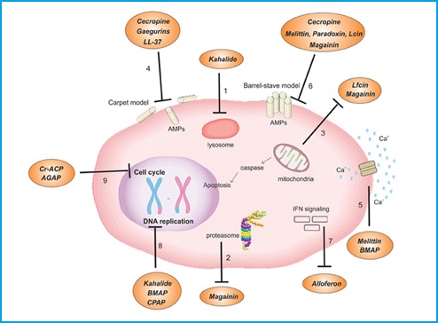
(1) Modification of the lysosome membrane leading to an acidification of the intracellular environment and cell death. (2) Amplification of the proteasome activity. (3) Induction of mitochondrial pathway of apoptosis by either the cytochrome c release into the cytoplasm or activation of the caspase cascade. (4) Pore formation by the carpet model. (5) Increase of the influx of Ca2+. (6) Formation of pore by either the toroidal or the barrel-stave models. (7) Activation of an immune modulatory pathway by induction of NK and IFN. (8) Inhibition of genes involved in DNA replication. (9) Arrest of cell cycle G0, G1, or S phases. Lfcin, lactoferrcin; BMAP, bovine myeloid antimicrobial peptide; Cr-AMP, Chlorella pyrenoidosa antitumor polypeptide.
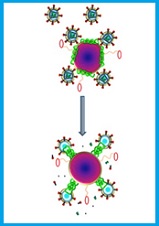
Cells in several types of cancer, such as renal, lung, liver, prostate, bladder, breast, and leukemia, can be targeted by melittin. It is well-known that melittin is a natural detergent with the capacity to form tetramer aggregates on membranes, which lead to disorders in the structure of phospholipid bilayers, changes in membrane potential, aggregation of membrane proteins, as well as the induction of hormone secretion. Furthermore, this membrane disruption directly or indirectly leads to alterations in enzymatic systems, such as G-protein, protein kinase C, adenylate cyclase, and phospholipase A. Melittin can even inhibit calmodulin, a calcium-binding protein that plays a crucial role in cell proliferation. Tumoral cells expose anionic phospholipids, mainly phosphatidylserine, on the external leaflet of the plasma membrane, and this feature can allow the preferential binding of cationic peptides, like melittin, relative to normal cells. Melittin studies with numerous types of cancer cells and in vivo animal models have demonstrated its antiproliferative activity. Furthermore, recent studies have demonstrated that melittin has anti-angiogenesis properties.
However, when a therapeutic dose of melittin is injected in vivo, some side effects, such as liver injury and hemolysis, were observed. To minimize these emerging lesions in off-target tissues, the following three strategies have been designed: (1) conjugaton of melittin to an antibody or a targeting component; (2) development of shielded pro-cytolytic melittin systems; and (3) synthesis of melittin-transporting carriers.
With regard to the first approach, a melittin-based recombinant immunotoxin obtained by fusion of genes that encoded an antibody fragment derived from the murine monoclonal antibody K121 with an oligonucleotide encoding melittin was tested successfully in vitro. Another study was based on a recombinant immunotoxin of melittin fused to an anti-asialoglycoprotein receptor (ASGPR) single-chain variable fragment antibody (Ca) which conferred targeting and ASGPR-specific cytotoxicity to hepatocellular carcinoma cells. Finally, a recent study characterized a CTLA-4-targeted scFv-melittin fusion protein as a potential immunosuppressive agent for organ transplant. In this regard, the selective cytotoxicity of the peptide construction was confirmed in preliminary biological activity assays.
Related to the pro-cytolytic melittin, by taking advantage of tumor matrix metalloproteinase 2 (MMP2) overexpressed on cancer cell membranes, an MMP2 cleavable melittin/avidin conjugate was built. Melittin coupled to avidin becomes inactive, but when released from the conjugate it induces immediate cell lysis. A similar idea was published years later, this time using avadin, the latency-associated peptide (LAP) domain of the transforming growth factor beta (TGF-β). In this approach, LAP dimerization conferred latency to the MMP2-cleavable melittin-LAP fusion protein.
Regarding pro-cytotoxic melittin systems, a design was based on the mixture of melittin with the anionic detergent sodium dodecyl sulfate formulated into poly(D,L-lactide-co-glycolide acid) nanoparticles by an emulsion solvent diffusion method. The inhibitory in vitro effects of these 130 nm-diameter melittin-loaded nanoparticles on breast cancer MCF-7 cells were promising. Another interesting carrier was a pegylated immunoliposome coupled to a humanized antihepatocarcinoma single-chain antibody variable region fragment and loaded with a bee venom peptide fraction. A similar pegylated immunoliposome but using only melittin as cargo and the complete antibody trastuzumab as targeting component was designed to combat HER2-overexpressing human breast cancer cell lines. The three aforementioned nanoparticles are not suitable for systemic administration because melittin can be released in blood vessels during transport, particularly in liposomes, which can be disrupted by the lytic peptide. To overcome this drawback, Samuel A. Wickline’s group developed a perfluorocarbon nanoemulsion vehicle incorporating melittin into its outer lipid monolayer. This nanocarrier of approximately 270 nm in diameter presented favorable pharmacokinetics, accumulating melittin in murine tumors in vivo and causing a dramatic reduction in tumor growth without any apparent signs of toxicity. Finally, the most recent ultra-small diameter melittin-nanoparticle (<40 nm) successfully tested in vivo with few side effects is the patented α-melittin-NP. This nanoparticle comprise 1,2 dimyristoyl-sn-glycero-3-phosphatidylcholine (DMPC) decorated with the hybrid peptide formed by peptide D-4F and melittin via a GSG linker, the peptide D-4F being a peptide that mimics a high-density lipoprotein (HDL).
[1] Mulder Kelly, Lima Loiane Alves, Miranda Vivian, Dias Simoni, Franco Octavio. Current scenario of peptide-based drugs: the key roles of cationic antitumor and antiviral peptides. Frontiers in Microbiology (2013) DOI: 10.3389/fmicb.2013.00321
Endosomolytic Properties
The strategy of packing and carrying small interference RNA (siRNA) using a wide variety of systems for gene therapy has been increasingly followed in recent years. The efficiency mediated by these drug delivery systems is strongly dependent on their endosomal escape capability, otherwise the siRNA would be degraded in endolysosomes. One mechanism designed for endosomal release is the use of fusogenic peptides, which are generally short amphipathic sequences between 20 and 30 amino acids in length and capable of disrupting biological membranes at endosomal pH. One of the first highly innovative studies using melittin consisted of reversibly masking the membrane-active peptide using maleic anhydride derivative. At neutral pH, the lysine residues of melittin were covalently acylated with anhydride, thereby inhibiting the membrane disruption activity of the peptide. Under acidic conditions such as those present within endosomes, the amide bond of the maleamate was cleaved, thus unmasking melittin. Similar studies performed by Ernest Wagner et al. showed that melittin analogs with high lytic activity at acid pH enhance the transfection of oligonucleotides in cell cultures and in in vivo mouse models. Very recently, a derivative of melittin (p5RHH) was reported to successfully trigger siRNA release into the cellular cytoplasm. The company Arrowhead Therapeutics is currently developing ARC-520 as a novel siRNA-based therapeutic to knock down the expression of viral RNAs of chronic hepatitis B virus. They describe the use of a coinjection of a hepatocyte-targeted, N-acetylgalactosamine-conjugated melittin-like peptide (NAG-MLP) with a liver-tropic cholesterol-conjugate siRNA (chol-siRNA) targeting coagulation factor VII. Preclinical studies with animals as well as Phase I assays have revealed that melittin promotes delivery without generating anti-melittin antibodies. In March 2014, Phase II trials of ARC-520 were started for patients with chronic hepatitis B virus.
Neurodegenerative Disorders
Use of Bee Venom and its components for the treatment of neurodegenerative diseases in in vivo models.[1]
|
Venom or Compound |
Neurological Disease |
Model Tested |
Administration via |
Dose |
|
Bee venom |
Parkinson’s Disease |
1-methyl-4-phenyl-1,2,4,5-tetrahydropyridine |
s.c. acupuncture (point GB34) |
0.02 mL bee venom (1:2000 w/v) |
|
once every 3 days for 2 |
||||
|
Bee venom |
Parkinson’s Disease |
MPTP in mice |
s.c. acupuncture (bilateral point ST36) |
A single injection (0.6 |
|
Bee venom |
Parkinson’s Disease |
MPTP/probenecid in mice |
i.p. |
Two injections 3.5 days |
|
Low—12 µg/kg/BW |
||||
|
High—120 µg/kg/BW |
||||
|
Bee venom |
Parkinson’s Disease |
MPTP in mice |
i.p. |
one i.p. injection |
|
BV (1 mg/kg) every day for 6 |
||||
|
Bee venom |
Parkinson’s Disease |
Rotenone-induced oxidative |
s.c. acupuncture (point GB34) |
0.02 mL bee venom (1:2000 w/v) |
|
once every 3 days for 2 |
||||
|
Bee venom |
Multiple Sclerosis |
Experimental allergic |
– |
2 mg/kg or 5 mg/kg |
|
Bee venom |
Amyotrophic Lateral Sclerosis |
hSOD1G93A transgenic |
s.c. acupuncture (bilateral point ST36) |
0.1 µg/g—3 times/week for 2 |
|
Bee venom |
Amyotrophic Lateral Sclerosis |
hSOD1G93A transgenic |
s.c. acupuncture (bilateral point ST36) i.p. |
0.1 µg/g—3 times/week for 2 |
|
Apamin |
Parkinson’s Disease |
MPTP/probenecid mice |
i.p. |
Two injections 3.5 days |
|
Low—0.5 µg/kg/BW |
||||
|
High—1.0 µg/kg/BW |
||||
|
Melittin |
Amyotrophic Lateral Sclerosis |
hSOD1G93A transgenic |
s.c. acupuncture (bilateral point ST36) |
0.1 µg/g twice a week |
The effect of Melittin on the CNS has been documented since 1973, when studies showed its marked effect on inhibiting general behavior, exploratory activity and “emotionality”, in addition to disrupting spontaneous and evoked bioelectric activity in the brain. Moreover, high doses of this peptide can induce a depressant effect evaluated by electroencephalography in anesthetized cats. This effect was associated with reduced systemic blood pressure.
In 2011, Yang and collaborators studied the therapeutic effect of Melittin in a transgenic mouse model for ALS. In this model, Melittin-treated animals exhibited a decline in the number of activated microglia and expression of proinflammatory factor TNF-α, inhibiting the increased neuroinflammation responsible for neuronal death in this disease. Moreover, Melittin regulates the production of misfolded proteins by activating chaperones and alleviating α-synuclein post-translational modification, an important mechanism for PD and ALS pathologies. Melittin also restored proteasome activity in the brainstem and spinal cord. Interestingly, treatment with this alkaline peptide in a symptomatic ALS animal model improved motor function and reduced neuronal death.
Additionally, in vitro assays revealed the potential in Melittin as an agent for the prevention of neurodegenerative diseases, considering its ability to inhibit the apoptotic factor and cell death in neuroblastoma SH-SY5Y cells. Melittin also demonstrated a potent suppressing effect on proinflammatory responses for BV2 microglia by reducing proinflammatory mediators and production of NO, PGE2 and cytokines. Thus, it is suggested that this compound may have significant therapeutic potential for the treatment of neurodegenerative diseases accompanied by microglial activation, such as PD
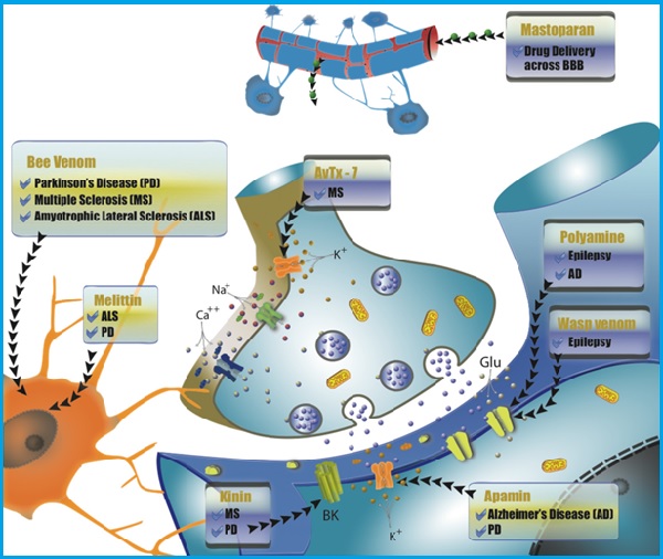
Main targets for wasp and bee venoms in the nervous system according to the type of neurodegenerative disorder treated.
Recently, Dantas and colleagues (2014) investigated the pharmacological effects of Melittin on the nervous system of mice. The animals were submitted to behavioral tests, including the catalepsy test, open field and apomorphine rotation tests. The results showed that mice treated with Melittin displayed no cataleptic effects or changes in motor activity, although there was a reduction in the effects induced by the apomorphine test. As such, the authors found that Melittin exhibited antipsychotic properties and may be an alternative for the treatment of psychotic diseases, reducing the classic side effects caused by conventional neuroleptic drugs.
[1] Toxins (Basel). 2015. DOI: 10.3390/toxins7083179 PMCID: PMC4549745 http://www.mdpi.com/2072-6651/7/8/3179/htm#table_body_display_toxins-07-03179-t001
Melittin, the Major Pain-Producing Substance of Bee Venom [1]
[1] Neurosci Bull 2016 Melittin, the Major Pain-Producing Substance of Bee Venom. Jun Chen, Su-Min Guan, Wei Sun, Han Fu. DOI: 10.1007/s12264-016-0024-y PMCID: PMC5563768